

If an intervention is being considered, any duplex ultrasound study should be followed by CTA or MRA, or another duplex ultrasound by a second operator. If carotid artery stenting is being considered, the duplex ultrasound should be followed by CTA or MRA in order to obtain more information on the anatomy of the aortic arch, as well as intra- and extracranial circulation. Table 3 shows that all modalities have similar performance in detecting a carotid artery stenosis.
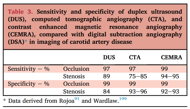

Both CTA and MRA have benefits and drawbacks. The benefit of CTA is that it is easily available in most settings, it is a quick examination that gives a lot of information about anatomy, additional stenosis in the proximal carotid artery or brachiocephalic artery, and it provides additional information on the contralateral carotid artery, vertebral arteries and the circle of Willis. However, especially in heavily calcified plaques, it can be harder to precisely calculate the degree of stenosis. MRA has the benefit that it is not disturbed by heavily calcified lesions, but it might be less available in most hospitals. Compared to duplex ultrasound, both modalities give additional anatomical information, such as tortuosity, tandem lesions, side branches, that can be taken into consideration when performing an intervention.

”Our

Below you can see an example of an MRA of the carotid arteries of another patient.

Lesson Content
Feedback E-Library
Give us your feedback about our E-Library !
How would you rate your experience?
Do you have any additional comment?
Submit
Thank you for submitting your feedback!
ESVS
Privacy Overview

This website uses cookies so that we can provide you with the best user experience possible. Cookie information is stored in your browser and performs functions such as recognising you when you return to our website and helping our team to understand which sections of the website you find most interesting and useful.